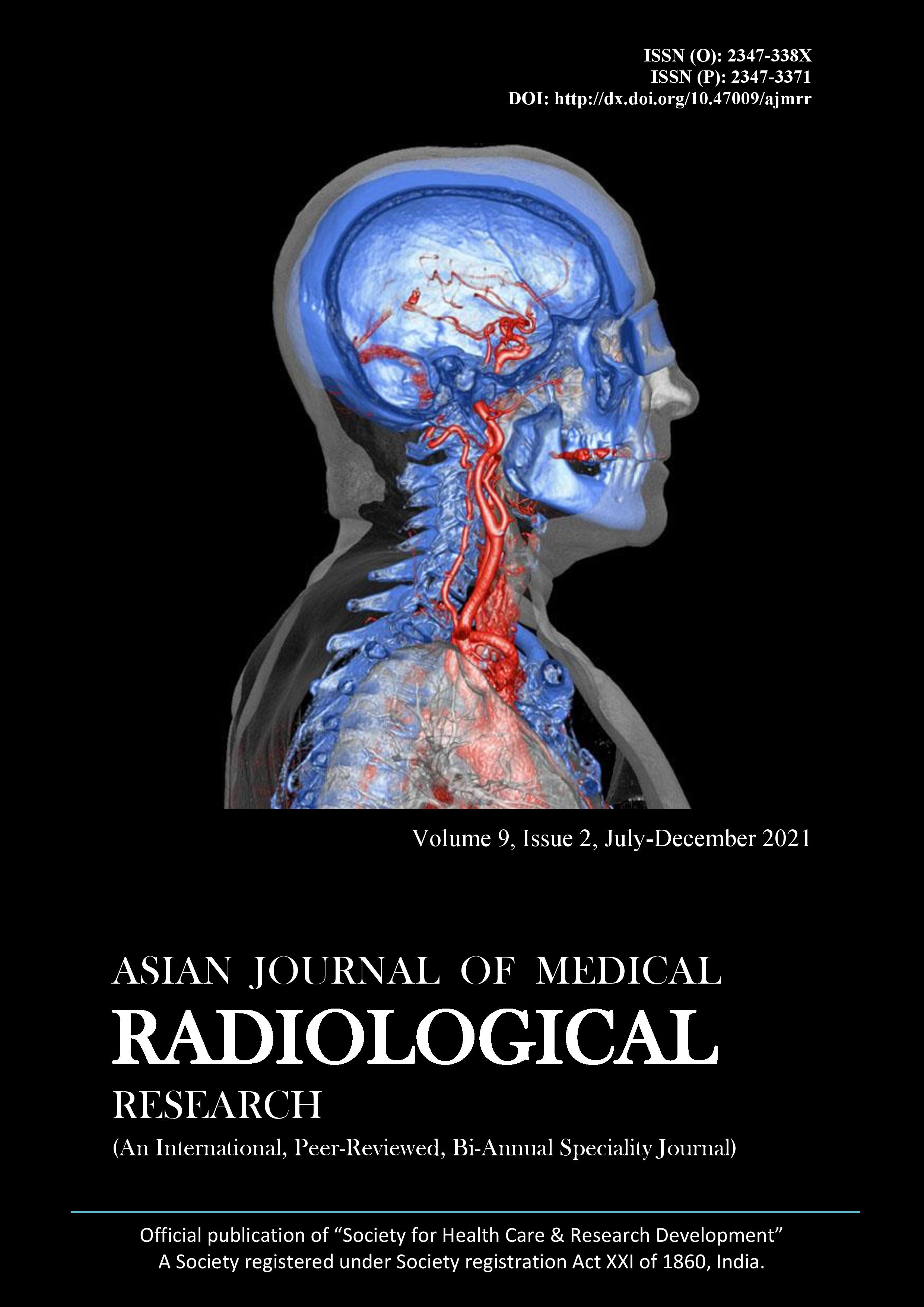
A Study of Circle of Willis and Normal Cerebral Circulation Variants on CT and MR Angiography
Circle of Willis and Normal Cerebral Circulation Variants on CT and MR Angiography
Abstract
Background: The Circle of Willis is a polygon constituting the anastomosis of the internal carotid and vertebral systems that permits cerebral arterial circulation. The main objective of this study was to assess the role of angiography in the evaluation of variant anatomy of Circle of Willis and to determine its relation with various associated vascular pathologies. Subjects and Methods: This Hospital-based prospective study was carried out over 2 years from July 2019 to August 2021 at the Department of Radiodiagnosis, Narayana medical college, Nellore. The study population included 200 patients- 130 cases were imaged on a 3 Tesla MRI scanner and the rest 70 on a 128- slice CT scanner. MIP and 3D-reformatted images were scrutinized to ascertain the ultimate configuration of the CW and the presence of vascular pathology. Results: Out of the two hundred cases, Complete and balanced CoW was appreciated in 27.9% of them. Posterior circulation variations were eyed in 43.8% and anterior circulation variations in 19.6%. Combined anterior and posterior circulation variations were noticed in 8.7 %. Hypoplastic/ aplastic Posterior communicating arteries were the most familiar in segmental variations. No immediate or prompt correlation was established between the attained results and associated vascular pathologies. Conclusion: The study results demonstrate slight differences in the CW configuration. A significant proportion of complete anterior CW was espied in female patients. Posterior circulation variants were the commonest among both men and women. No remarkable association was revealed between CW configuration and the occurrence of aneurysms/AVMs in this analysis. Normal variants of the cerebral circulation are customary, and most such anomalies can be identified at CT and MR angiography.
Downloads
Copyright (c) 2021 Author

This work is licensed under a Creative Commons Attribution 4.0 International License.


